Multicellular organisms comprise millions to trillions of cells that work together in harmony. The cells in various tissues have unique shapes and carry out different roles for the organism. Despite their varied functions, each somatic cell typically contains the same chromosomes and they share the same genetic information. All the cells in a fully developed organism trace back to a single cell formed when the parents’ male and female gametes combined. This initial cell marked the beginning of the organism’s life.
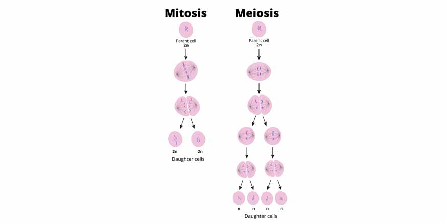
Cell division is when a single parent cell splits into two or more daughter cells. This process is typically part of a bigger cycle known as the cell cycle. The cell’s nucleus divides during cell division, and the DNA is replicated. There are two main types of cell division: mitosis and meiosis. When people talk about “cell division,” they usually mean mitosis, which is how new body cells are formed. On the other hand, meiosis is the division that produces cells, like eggs and sperm. Mitosis is a fundamental process in life.
Understanding mitosis and meiosis difference
|
Mitosis |
Meiosis |
|
Discovered by Walther Flemming |
Discovered by Oscar Hertwig |
|
This type of cell division occurs in all types of cells, even sex cells |
This type of cell division occurs in meiocytes (specialised cells) |
|
Mitosis occurs in somatic cells |
Meiosis occurs in germ cells |
|
Prophase, metaphase, anaphase and telophase are the stages of mitosis |
Different stages of meiosis are Prophase I, metaphase I, anaphase I, and telophase I and then enter into Meiosis II which includes the following phases Prophase II and then metaphase II, anaphase II, and telophase II. |
|
Only one nuclear division occurs |
Two nuclear divisions occur |
|
Chromosome number remains unaffected |
The chromosome number is reduced to half |
|
Mode of reproduction- Asexual |
Sexual |
|
Cytokinesis occurs in telophase |
Cytokinesis occurs in Telophase I and II |
|
No recombination. |
Recombination and crossing over take place between homologous chromosomes. |
Stages of Mitosis and Meiosis
Mitosis
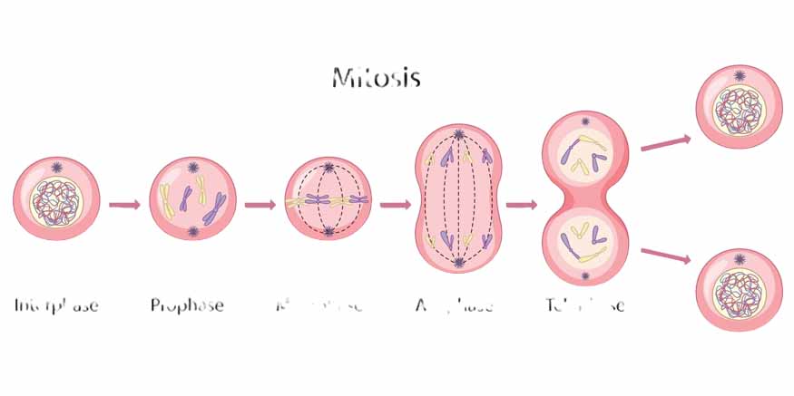
Prophase: In prophase, the chromatin starts to tighten up and forms visible chromosomes. At the same time, the mitotic spindle begins to develop, and the nuclear envelope begins to disintegrate, which allows the spindle fibres to connect with the chromosomes.
Metaphase: During metaphase, the chromosomes line up along the centre of the cell, called the metaphase plate. The spindle fibres attach to the centromeres of the chromosomes, making sure that each new daughter cell will get an exact copy of the chromosomes.
Anaphase: Anaphase is when the sister chromatids are pulled apart. The spindle fibres get shorter, dragging the chromatids to opposite ends of the cell, which guarantees that each daughter cell will have the same set of chromosomes.
Telophase: In telophase, the chromosomes arrive at the poles of the cell, and the nuclear envelope starts to reform around each group of chromosomes, creating two separate nuclei. The chromosomes then begin to loosen back into chromatin.
Cytokinesis: Cytokinesis is the last step, where the cytoplasm splits, leading to the formation of two individual daughter cells. This process makes sure that each daughter cell has all the necessary parts to live and work on its own.
Meiosis
Meiosis involves two main divisions: meiosis I and meiosis II, each containing specific stages.
Meiosis I:
Prophase I: During this phase, homologous chromosomes come together and exchange genetic information through crossing over. The chromatin becomes tightly packed into visible chromosomes, the nuclear envelope disintegrates, and the spindle apparatus begins to form.
Metaphase I: Here, the homologous chromosome pairs line up along the metaphase plate. Unlike mitosis, where single chromosomes align, in meiosis I focus on aligning these pairs, ensuring that each daughter cell gets one chromosome from each pair.
Anaphase I: The homologous chromosomes are pulled apart and moved to opposite ends of the cell. This division reduces the chromosome number, making sure each daughter cell ends up with a haploid set.
Telophase I: The chromosomes arrive at the poles, and the nuclear envelope may start to reform. Cytokinesis then occurs, leading to the formation of two haploid daughter cells.
Meiosis II:
Prophase II: In this stage, the chromatin condenses into visible chromosomes again, the nuclear envelope breaks down, and the spindle apparatus forms in each of the two daughter cells produced from meiosis I.
Metaphase II: The chromosomes line up at the metaphase plate, similar to what happens in mitosis, but this time in a haploid setting.
Anaphase II: The sister chromatids are separated and pulled to opposite ends of the cells. This division is equational, ensuring that each of the four resulting daughter cells has a unique combination of chromosomes.
Telophase II: The chromatids reach the poles and the nuclear envelope reforms around each set. Cytokinesis then occurs, resulting in four genetically distinct haploid daughter cells.
Mitosis and meiosis are essential mechanisms of cell division that serve distinct yet equally important roles in biology. Mitosis ensures growth, repair, and asexual reproduction through the production of genetically identical cells. In contrast, meiosis generates genetic diversity and enables sexual reproduction by producing haploid gametes. Understanding the mitosis and meiosis difference, along with the stages of mitosis and meiosis, provides a comprehensive insight into the complexity and beauty of life at the cellular level.
At Center Point School, we strive to impart a profound understanding of these fundamental biological processes, ensuring our students are well-equipped with the knowledge and skills to appreciate the intricacies of life. Our holistic educational approach fosters curiosity and critical thinking, preparing learners for future scientific endeavours and discoveries.

